Introduction: Epstein-Barr virus (EBV) positive NK/T cell lymphoma is a subtype of non-Hodgkin's lymphoma strongly associated with EBV infection. EBV infection has been reported in 85-100% of cases. It has been suggested to be central in pathogenesis. The incidence of EBV+ NK/T cell lymphoma is increasing worldwide, particularly in Asia and Latin America, and it has a poor prognosis with few effective treatment options. Therefore, novel approaches for treating EBV-associated NK/T cell lymphoma are urgently needed. Developing chimeric antigen receptor (CAR)-T cells therapy targeting EBV specific protein in the context of this EBV-positive aggressive lymphoma will provide a novel therapeutic strategy for patients with limited treatment options. Targeting EBV lytic protein for control EBV in vivo replication and tumorigenesis have been explored lately. The gp350 protein is a major glycoprotein on the surface of EBV during its lytic phase, after fusion between virus and host cell membrane, gp350 is left on the host cell surface. The distinct surface distribution pattern of gp350 in both freshly infected host cells and the lytic phase host cells renders it an attractive target for immunotherapy.
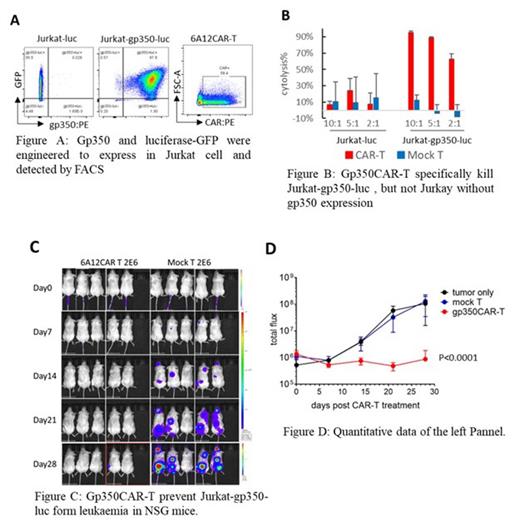
Method and Result: We developed EBV specific CAR-T cells targeting gp350. We utilized single-chain variable fragments (scFvs) derived from four novel, gp350-binding, neutralizing monoclonal antibodies (mAbs) (12C10, 7D11, 6A12, 7A8) to generate the CAR constructs. We demonstrated that these scFvs, when fused to a second-generation CAR backbone consisting of 4-1BB and CD3ζ signal domains, are efficiently expressed on the surface of transduced T cells. These gp350 CAR-T cells exhibit specific activation and cytotoxic effects in vitro. Of note, the clone 6A12 scFv of CAR displayed the most potent cytotoxicity effects in gp350 positive cancer coculture toxicity assays. To optimize the CAR structure further, we designed various linker and transmembrane domains based on the clone 6A12 scFv, including CD8 linker, IgG4 hinge, IgG1 Fc linker, and CD28 linker and transmembrane domains. Our cytotoxicity assay screening indicated that the CD8 linker and transmembrane domain resulted in the most advanced cytotoxicity and target specificity (Figure A, B). To assess gp350 CAR-T efficacy in vivo, we established a T-cell leukaemia animal model in NOD immune-deficient mice through intravenous injection of Jurkat-gp350-luc cells. On post xenograft day 7, the Jurkat-gp350-luc xenografted mice were separated into three groups and each group with 5 mice (except PBS group with 2 mice). CAR-T and T cell were infused by tail vein injection with mock T 2x10^6/mouse, 6A12CAR-T 2x10^6/mouse and PBS control. The tumor growth in mice was monitored by in vivo luminescence with IVIS imaging system. Upon engraftment, Jurkat-gp350-luc cells established disseminated leukaemia expanding preferentially in the spine, femur, head, and pelvis, with few cells in peripheral blood. Mice receiving control mock T cells or without T cell PBS control developed rapid disease progression and were euthanized by day 35 after Jurkat-gp350-luc engraftment. In contrast, mice receiving gp350 CART cells were significantly protected from rapid progression (Figure C, D).
Conclusion: In pre-clinical development, we designed, constructed, and screened gp350 CAR-T cells to demonstrate its efficacy in vitro and in vivo against and kill gp350 positive leukaemia cells and prevent formation and development leukaemia in xenograft mice model.
Disclosures
No relevant conflicts of interest to declare.


This feature is available to Subscribers Only
Sign In or Create an Account Close Modal